
✓
No cell block
Direct-to-slide cryofixation removes embedding, sectioning, and repeated section consumption from the core workflow.
The NGC workflow replaces cell-block preparation and sequential IHC with a single-slide cytology process: cytocentrifugation, cryofixation, eight-channel staining, and hybrid digital review.
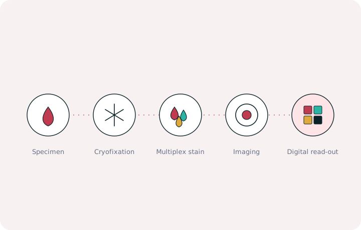
From sample to slide in under 4 hours.
Native or EDTA-collected sample arrives in standard cytology containers.
Cells are concentrated and deposited directly onto a microscope slide.
Cryofixator™ preserves morphology and nucleic acids without cell blocking.
Cryostainer™ performs multiplex IF with seven antibodies plus nuclear counterstain.
Selected areas are digitized for side-by-side channel and merged-image review.
37% Less manual handling. 75% Fewer touch points. 95% Less sample preparation time.
Direct-to-slide cryofixation removes embedding, sectioning, and repeated section consumption from the core workflow.
The consolidated workflow reduces manual sample movement and keeps fixation, staining, and review tightly coordinated.
Up to eight data points are generated on a single slide instead of distributing markers across serial slides.
The performance page summarizes turnaround, handling, data yield, and benchmark comparisons from the deck.