
▽
Cryofixation™ hardware
Controlled cryo-environment processing preserves cellular morphology and antigen integrity directly on the slide — eliminating the cell-block dependency.
Direct-to-slide cryofixation, multiplex immunostaining, and digital review — verified at the bench and validated in independent clinical studies.
Regulatory status: Cryosuite platform, Cryophore™ multiplex panels, and their reagents are EU-IVDR registered (Class A). Specific panel regulatory status varies by market — please contact us for current documentation.
A cytology-first platform architecture for routine use. Same-day, multi-biomarker diagnostics on minimally invasive samples — without cell-block preparation or sequential staining.
Controlled cryo-environment processing preserves cellular morphology and antigen integrity directly on the slide — eliminating the cell-block dependency.
Proprietary fluorophore-conjugated antibody chemistry engineered for 8-channel simultaneous staining with minimal spectral crosstalk.
Pre-configured, expert-vetted panel compositions covering effusion and FNA cytology. Pleural Fluid 1/2/3, Ascitic Fluid, and a growing pipeline.
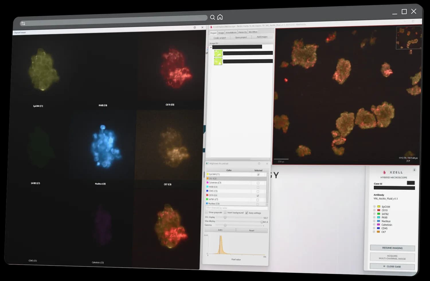
Sub-30-second slide digitisation, multi-channel single-cell viewer, on/off channel toggling, customisable merge view. AI-ready architecture.
A linear, single-pass workflow that replaces multi-day serial IHC iteration with same-day multiplex evaluation.
Effusion or FNA cytology specimens. Wet monolayers, smears, imprints, or frozen sections — upstream-agnostic. No cell-block preparation required.
Controlled cryo-environment preserves morphology and antigen integrity. Minimises non-specific binding. Sample remains compatible with downstream NGS.
Single-pass, 8-channel simultaneous immunofluorescence using the chosen Cryophore™ panel. No iterative re-staining cycle.
Sub-30-second whole-slide digitisation. Multi-channel single-cell viewer with on/off toggling and merge views. Read-out accessible from any workstation.
Designed to support standardised cytology workflows where every biomarker counts and sample material is scarce.
8 biomarkers from a single slide in under four hours — replacing 24-48-hour single-marker cycles.
100 % of the remaining specimen is preserved for downstream NGS and molecular profiling. No iterative depletion.
Cytology-first design — not an off-label workaround of histology IHC. EU-IVDR registered platform and Class A panels.
Direct-to-slide cryofixation removes the upstream cell-blocking step that wastes time and material in conventional workflows.
Expert-vetted Cryophore™ panels deliver reproducible multiplex performance across labs and operators. No bespoke validation per case.
Single-cell viewer with channel-level controls. 30 000+ manually annotated cells form an AI-ready dataset for image-assisted interpretation.
Reference: cell-block + sequential immunohistochemistry / immunocytochemistry, the prevailing multi-marker workflow.
1 biomarker per slide · 24-48+ hours per iteration · material depletion at every step · manual microscopy review · non-standardised workaround for cytology · IVDR-restricted off-label use.
8 biomarkers per slide · 3-4 hours end-to-end · minimal material consumption · digital multi-channel review · cytology-first architecture · EU-IVDR registered (Class A) platform + panels.
Comparative metrics from independent evaluations. Reference workflow: cell-block + immunohistochemistry / immunocytochemistry.
+112% improvement in detection sensitivity vs the reference workflow.
−92% reduction in time-to-result, enabling same-day clinical decisions.
−49% less laboratory hands-on time per case.
+700% biomarker data extracted from a single specimen — 8-channel multiplex.
+64% more true-positive identifications across the evaluated cohort.
−100% non-diagnostic cases. No first-pass diagnostic ambiguity in this cohort.
Every antibody entering the X-ZELL portfolio survives a multi-stage wet-lab and computational verification process.
Commercially available clones evaluated for relevance, specificity, and multiplex compatibility.
Survived wet-lab stress-testing and computational cross-checks. Ready for clinical use.
Verified zero cross-reactivity across the qualified portfolio.
Clean, unambiguous results in every multiplex panel configuration.
Real-world validation across 246 mixed effusion and FNA specimens at two independent German cytopathology institutes, under IVDR standards.
Routine effusion and FNA specimens collected at two independent cytopathology institutes in Germany.
~22% baseline QC exclusion — a real-world figure reflecting routine FNA imprint variability, not a laboratory ideal.
Review the current validation report, specimen context, and comparator method before using performance statements in evaluation or implementation decisions.
Intra-operative fine-needle workflow: the X-ZELL platform replaces single-marker, 24-hour ICC turnaround with same-day 7-channel multiplex.
Using the Cryophore™ Pleural Fluid 1 and Pleural Fluid 2 panels in routine intra-operative use.
Multi-channel digital evaluation accessible from any workstation, replacing manual microscopy review.
Eliminates the iterative repeat-staining loop; no repeat procedures across evaluated cases.
Beta evaluation and clinical validation at independent academic and clinical institutes across Germany, Croatia and Slovenia.
Peer-reviewed publications and international conference contributions covering platform chemistry, panel design, and clinical performance.
Manually annotated single-cell dataset forming the AI-ready foundation for image-assisted interpretation.
Independent international expert committee — multiple WHO guideline contributors, on the Cryophore™ panel design board.
Miceska, S., Kholová, I., & Kloboves Prevodnik, V. (2026). Acta Cytologica, 1–28. DOI: 10.1159/000550736
Bhakdi, S. C., Walch, D., Fives, C., Grote, S., Hunfeld, K. P., & Battmann, A. (2025). 22nd International Congress of Cytology, 1–94.
Bhakdi, S. C., & Battmann, A. (2023). Cytopathology, 34(S1), 5–55. DOI: 10.1111/cyt.13293
Bhakdi, S. C., & Thaicharoen, P. (2018). Methods and Protocols, 1(2), 1–11. DOI: 10.3390/mps1020020
Grote et al. (2026). Manuscript submitted. Operational metrics & diagnostic performance vs. cell-block + ICC/IHC.
Battmann et al. (2026). Data evaluation — diagnostic concordance and true-positive yield.
Quality management system certified. Manufacturing under formal QMS, with lot-level traceability per batch.
Cryosuite hardware + software + Cryophore™ panels and reagents are EU-IVDR registered (Class A).
Hardware, software and consumables are FDA-registered for the US market. International IP and EUDAMED registration in place.
A concise evidence layer for cytology laboratories evaluating multiplex cryoimmunostaining. Details should be interpreted in the intended specimen and regulatory context.
Peer-reviewed preliminary evaluation of multiplex cryoimmunostaining on routinely prepared cytospin samples from fine-needle aspiration biopsies and effusions.
Read article →Conference abstract listed in X-ZELL resources, relevant to effusion cytology workflows and routine-laboratory evaluation.
View resources →Public X-ZELL news describes interim German study data comparing NGC with conventional cell-blocking and IHC approaches for minimally invasive samples.
Read summary →Foundational publication referenced by X-ZELL for multi-channel fluorescence detection, relevant to the technical rationale for multiplex review.
See literature list →Shortcuts for teams evaluating workflow fit, panel availability or technical documentation.
A concise visual overview of the platform, intended as a first-pass resource for internal evaluation.
Open brochure →Definitions for Cryoimmunostaining™, Cryophore™, Hybrid Microscope and Next-Generation Cytology.
View glossary →Product availability, intended use and regulatory status vary by market. Request current documentation before procurement.
Request dossier →Our scientific liaison team can provide the complete analytical and clinical validation data, peer-reviewed publications, and IVDR documentation under NDA for qualified institutions.